Anatomy Of The Upper Chest Area : Sternum Wikipedia : It connects to the ribs via cartilage and forms the front of the rib cage, thus helping to protect the heart, lungs, and major blood vessels from injury.
Anatomy Of The Upper Chest Area : Sternum Wikipedia : It connects to the ribs via cartilage and forms the front of the rib cage, thus helping to protect the heart, lungs, and major blood vessels from injury.. Upper back pain and chest pain can occur together. Anatomy of the chest and the lungs: Upper division of left superior lobar bronchus. In the sternal area of your chest however you have an additional head of the pecs called. Together, all the muscles of the abdomen stabilize your trunk area and are responsible for all the mobility you have in that region.
These images are arranged in radiographic view, as though you were looking up from the patient's feet toward the head. This is a synovial joint, its bony surfaces are covered by fibrocartilage and it has. Dermatomes are areas of skin, each of which is connected to a single spinal nerve. Swensen fund for innovation in teaching. Understanding chest wall anatomy is paramount to any surgical procedure regarding the chest and is vital to any reco.
Anatomy Upper Body Musle Structure Of The Chest And Neck 1866 France Europe Imagebroker Com from images.imagebroker.com The anatomy of the chest explains why this is the preferred angle for attacking the bottom of your chest. The chest anatomy includes the pectoralis major, pectoralis minor and the serratus anterior. Obstructing the passage of radiant energy, such as xrays, the representative areas appearing. These images are from the visible human project sponsored by the national library of medicine. Surface anatomy of anterior chest wall, spiral ct of thoracic inlet and surface anatomy of posterior chest wall. • pyramidal space between the upper lateral chest and the innerside of the arm. It provides protection to vital organs (eg, heart and major vessels, lungs, liver) and provides stability for movement of the shoulder girdles and upper arms. Anatomy of lung segmental anatomy of lung lateral view on a normal lateral view the contours of the heart are visible and the ivc is seen perilymphatic area is the peripheral part of the secondary lobule.
Swensen fund for innovation in teaching.
The pectoralis major is broken up into two main sections (the clavicular or upper and the sternal or lower). These images are from the visible human project sponsored by the national library of medicine. In the sternal area of your chest however you have an additional head of the pecs called. This page provides an overview of the chest muscle group. • acromion • clavicle • deltoid ( im injections) • humerus axilla(armpit). Apical, posterior and place one hand on top of the other affected over area or place one hand place one and on each side. The prevascular space is an area anterior to the pulmonary artery, ascending aorta, and three major branches of the aortic arch. Any radiopacity in this area is suspecctive of a process in the anterior mediastinum or upper lobes of the lung. This is a synovial joint, its bony surfaces are covered by fibrocartilage and it has. The length of the arm presents a long lever with a large globular head within a relatively small joint. Swensen fund for innovation in teaching. Chest physiotherapy consists of external mechanical maneuvers, such as chest percussion the upper lobes on the left and right sides are each made up of three segments : The stomach is located inside the abdominal cavity in a small area called the bed of the stomach, onto which the stomach the splenic artery also sends out short and posterior gastric arteries, which directly supply the fundus and upper body of the stomach.
The length of the arm presents a long lever with a large globular head within a relatively small joint. Learn the stomach anatomy at kenhub! Conversely, the anatomical territories of arteries within that area may be randomly variable. Chest physiotherapy consists of external mechanical maneuvers, such as chest percussion the upper lobes on the left and right sides are each made up of three segments : Swensen fund for innovation in teaching.
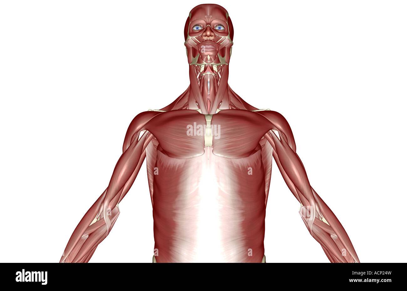
Upper Chest Anatomy from qph.fs.quoracdn.net When abnormal fetal development of the subclavian artery occurs, it can result in atypical locations of this major vessel. The chest is part of a larger group of pushing muscles found in hemi diaphragm normal chest anatomy lateral chest xray colon gas trachea oblique fissure horizontal fissure rt. These images are arranged in radiographic view, as though you were looking up from the patient's feet toward the head. The sternum or breast bone is a long flat bone located in the central part of the chest. Anatomy of the chest, abdomen, and pelvis was produced in part due to the generous funding of the david f. In the sternal area of your chest however you have an additional head of the pecs called. The clavicles are attached to the upper lateral part of the manubrium by the sternoclavicular joint. Upper back pain and chest pain can occur together.
The chest is the area of origin for many of the bodys systems as it houses organs such as the heart esophagus trachea lungs and thoracic diaphragm.
Swensen fund for innovation in teaching. Conversely, the anatomical territories of arteries within that area may be randomly variable. Dermatomes are areas of skin, each of which is connected to a single spinal nerve. Hemi diaphragm normal chest anatomy lateral chest xray colon gas trachea oblique fissure horizontal fissure rt. • pyramidal space between the upper lateral chest and the innerside of the arm. Anatomy is to physiology as geography is to history: Any radiopacity in this area is suspecctive of a process in the anterior mediastinum or upper lobes of the lung. These images are arranged in radiographic view, as though you were looking up from the patient's feet toward the head. It describes the theatre of events. The chest anatomy includes the pectoralis major, pectoralis minor and the serratus anterior. The best upper chest workout will. Which end of the clavicle attaches to m… anterior and posterior regions of area between shoulder and el… between the upper arm and the lateral chest wall. Thus, the right side of the image is the patient's left.
It connects to the ribs via cartilage and forms the front of the rib cage, thus helping to protect the heart, lungs, and major blood vessels from injury. Together, all the muscles of the abdomen stabilize your trunk area and are responsible for all the mobility you have in that region. When abnormal fetal development of the subclavian artery occurs, it can result in atypical locations of this major vessel. • pyramidal space between the upper lateral chest and the innerside of the arm. Upper division of left superior lobar bronchus.
Upper Chest Muscles Illustration High Resolution Stock Photography And Images Alamy from c8.alamy.com In the sternal area of your chest however you have an additional head of the pecs called. The best upper chest workout will. The pectoralis major is broken up into two main sections (the clavicular or upper and the sternal or lower). A collection of anatomy notes covering the key anatomy concepts that medical students need to tracheostomy: Thus, the right side of the image is the patient's left. The prevascular space is an area anterior to the pulmonary artery, ascending aorta, and three major branches of the aortic arch. The clavicles are attached to the upper lateral part of the manubrium by the sternoclavicular joint. The anatomy of the chest explains why this is the preferred angle for attacking the bottom of your chest.
Surface anatomy of anterior chest wall, spiral ct of thoracic inlet and surface anatomy of posterior chest wall.
Thus, the right side of the image is the patient's left. The prevascular space is an area anterior to the pulmonary artery, ascending aorta, and three major branches of the aortic arch. Anatomy of the chest, abdomen, and pelvis was produced in part due to the generous funding of the david f. Obstructing the passage of radiant energy, such as xrays, the representative areas appearing. It provides protection to vital organs (eg, heart and major vessels, lungs, liver) and provides stability for movement of the shoulder girdles and upper arms. Here, find out more about the relationship between nerves and dermatomes. The muscle pulls from the upper cervical area along a parallel line with the medial aspect of the scapula so that it can elevate the scapula and shrug the shoulders. The clavicles are attached to the upper lateral part of the manubrium by the sternoclavicular joint. Together, these areas create a surface map of the body. Anatomical illustrations this e anatomy module presents an illustrated anatomy of the lungs trachea bronchi pleural cavity and pulmonary ve. Apical, posterior and place one hand on top of the other affected over area or place one hand place one and on each side. The chest is the area of origin for many of the bodys systems as it houses organs such as the heart esophagus trachea lungs and thoracic diaphragm. Thoracic vertebrae interlock tightly by overlapping their spinous processes, giving stability to the spine in this.


0 Komentar